Analysis of Scans
MRI of Knee
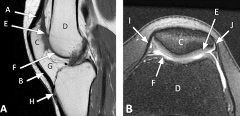
This is an MRI scan of the extensor mechanism of the knee. Figure 'A' shows the side view of the knee, while figure 'B' shows the knee from a bird's eye view. The arrow A pointing towards a tendon. Arrow B is also a tendon called the Patellar. Area C is where the Patella is and area D is the fenur. Arrows E and F are cartilage muscles that help support the knee. Arrow H shows an insertion of the tendon. Knees have mucles tissues which allows them to help us move since the main function of muscle tissues is for movement.
MRI of Shoulder
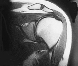
This image is a MRI scan of a shoulder. Shoulders have a lot of muscle tissues to help move our arms so by knowing that I believe that the darker parts in this scan show muscles and bones while the light parts are hollow structures or the protective barriers. The large circular light spot on edge of the shoulder could be a bone since that is the most important part of the shoulder; it helps move the arm. The large spot cannot be a hollow structure because a hollow structure will not be able to provide a lot energy for the movement of the arm. Along with the bones there also has to be a cartilage to provide structure, tendons to join bone to bone and liagments to join muscle to bone.
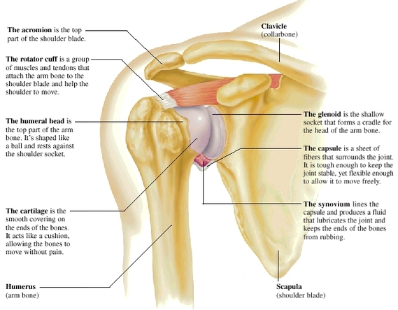
Parts of A Shoulder
You be the jugde! Did I get it right?
