How MRI Scans Work: The Parts
The most important part of an MRI scanner is the magnet, the “Magnetic” part in MRI. There is a long tube, the same one where the patient enters, that runs through a giant magnet from front to back. This tube is known as the “bore”. But the magnet used in MRI scanners isn’t like one of the magnets you stick on your kitchen fridge. These magnets are incredibly strong, and able to produce a large, stable magnetic field.
Most MRI scanners use a superconductive magnet, which consist of coils of wire through which a current of electricity is passed, creating a magnetic field. Maintaining such a large magnetic field requires a large amount of energy, which is created by superconductivity, which means reducing the resistance in the wires to almost zero. This is done by continuously bathing the wires in liquid helium at a freezing 452.4 degrees below Fahrenheit (or 269.1 below zero degrees Celsius). This cold is kept insulated by a vacuum. Superconductive magnets are very expensive, but the strong magnetic field they produce creates the highest-quality imaging. Other magnets can be used, such as Resistive magnets and Permanent magnets, but Resitive magnets, although they are structurally like a superconducting magnet, they lack the liquid helium which means they require a huge amount of electricity to work and are expensive to operate, while the latter is so heavy that it would be difficult to create one that could sustain a large magnetic field.
There are also three Gradient magnets in MRI scanners. These magnets have a much lower strength compared to the main magnet. While the main magnet is used to create and intense, stable magnetic field around the patient, the Gradient magnets create smaller, variable fields which allow different parts of the body to be scanned.
Another part of the MRI system is a set of coils that transmit a radiofrequency waves into the patient’s body. There is a different set of coils for different parts of the body, such as the knees, shoulders, neck and so on. These coils either conform to the body part being imaged, or are positioned very close to it during the exam. Other parts of the machine include an extremely powerful computer system and a patient table, which slides the patient into the bore.
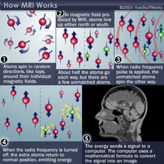
How hydrogen atoms react with the magnetic fields to form an image.
How MRI Scans
Works: The Procedure
The patient is stripped of all metal articles and credit
cards, or anything else that includes magnetic encoding (because when exposed
to the magnetic field, all information will be erased), and is placed on the
patient table, which then slides into the bore.
Depending on what parts need examining, the patient will either go head
or feet first. Once the body is at the exact center of the magnetic field
(known as the isocenter), the scan can begin.
When the patient slides into the bore, they are taking with them billions of atoms that make up the human body. Why is this so important, you ask? Because that is what the MRI measures to develop the scans. Atoms. In particular, the hydrogen atoms found in your body. Hydrogen atoms reside in soft tissues that are mostly made up of water or fat. These atoms randomly spin on their axis, and they all have a strong magnetic movement. This means that while they all go in various directions when in their natural state, but when introduced to a magnetic field, the atoms line up in the direction of the field. Since the magnetic field runs straight down the center of the machine, the hydrogen protons line up so they’re pointing either at the patients feet or head. About half the protons go each way, so most of the protons cancel each other out. So, for every atom that is lined up towards the feet, one is lined up towards the head. Only a few protons out of every million aren’t canceled out, meaning they have no pairs to cancel them out. And it’s these same unmatched atoms are what create the image.
Next, the MRI machine releases a radio frequency (RF) pulse that is specific to hydrogen. The system directs this pulse to the part of the body that is being examined. When the pulse is applied, the unmatched protons absorb the energy and resume spinning in different directions. This is the “Resonance” part of MRI. The RF pulse forces these atoms to spin in at a certain frequency and in a particular direction. This frequency is called the “Larmour frequency” and is calculated based on the tissue being imaged and the strength of the main magnetic field.
At more or less the same time, the three gradient magnets kick into action. These magnets are arranged in a certain way inside the main magnet so that when they’re turned on and off quickly in a specific way, they change the main magnetic field. This means that you can pick exactly what area you want an image of, known as the “slice”. These slices are as thin as a few millimeters and are extremely precise. They can be taken of any part of the body, in any direction, which is a huge advantage. Another advantage is that with the gradient magnets you can manipulate everything, so you don’t have to move for the machine to get an image in a different direction.
In the end of the scan, the RF pulse is turned off, and the hydrogen protons slowly return to their natural state within the magnetic field and release the energy they absorbed from the RF pulses. When they do this, they give off a signal that the coils pick up and sent to the computer system.
But how does the computer form an image? Well, the MRI scanner can pick out a point in the patient’s body, and essentially ask it “What sort of tissue are you?” The system goes through the patient’s body point by point, building up a map of tissue types as it goes along. Then, the system integrates all of this information to build up either 2-D images or 3-D models using a mathematical formula known as the “Fourier transformation”. The computer then receives the signal from the spinning protons as mathematical data and converts the data into a picture. This is the “Imaging” part of MRI.
The MRI system uses injectable contrast, or dyes, to alter the local magnetic field in the tissue being examined. The normal and abnormal tissues will react differently to this alteration, giving off different signals. When the signals are transferred to the image, the doctors can see different types of tissue abnormalities with more clarity than without contrast. An MRI system can display more than 250 shades of grey.
The process through which an MRI creates an image is long and complex, not to mention slightly uncomfortable for the patient. A tremendous amount of noise is produced, something like a continuous, rapid, hammering that is caused by the rising electrical current in the wires of the gradient magnets being opposed by the main magnetic field. This can be solved by wearing ear plugs or by listening to a music player. Another problem is that for a clear, high-quality image, the patient cannot move at all. MRI scans can take up to 20 to 90 minutes or more, and even very slight movement can cause distorted images and the process will have to be repeated. Also the exams are very expensive, as the equipment is too. However, despite a few draw backs MRI scanners are a big accomplishment in the world of biotechnology.
